
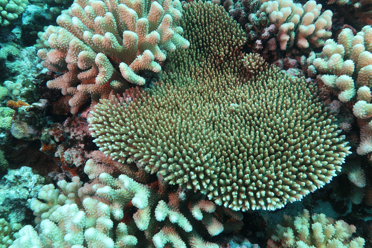
The branching coral, Acropora hyacinthus, is a prevalent coral species on the reefs of Mo’orea, French Polynesia, and was used as a focal species in this study.
Across the globe and in the Caribbean, coral reef ecosystems—home to an estimated 25 percent of all marine life—face crisis after crisis. Anthropogenic climate change leaves these delicate ecosystems increasingly vulnerable to stressors like mass bleaching, ultimately causing widespread declines in health.
In addition to these threats, stony coral tissue loss disease (SCTLD) has emerged and is decimating coral reef communities across the Caribbean at an alarming pace. Scientists are working to determine the cause of SCTLD, which was first observed off the Florida coast in 2014 and has since been reported in 28 Caribbean nations, to identify possible treatment methods and ways to better prevent its spread.
Bacteria and viruses have emerged as possible suspects for its pathogen, the disease-causing agent. Filamentous viruses in particular have gained recent attention as a possible contributor to the disease. However, researchers from the University of California, Berkeley and Rice University have discovered evidence that filamentous viruses may be globally distributed in corals, and not a component unique to this devastating disease.
Their new study, published today in The ISME Journal, probes the relationship between SCTLD and viral infections of dinoflagellates, a group of symbiotic algae crucial to the health of coral reefs.
“There are a lot of different hypotheses out there that try to identify the cause of this disease [SCTLD], but our work has clarified there might not be a ‘one pathogen-one disease’ model in play,” said Lauren Howe-Kerr, who led the research while a postdoctoral researcher at Rice University. “We will need more lines of evidence to determine what is causing this.”
Environmental Science, Policy, and Management professor Adrienne Correa, who joined Rausser College this fall from Rice University, served as the study’s senior author.
An emerging challenge
Coral colonies infected with SCTLD tend to exhibit rapidly expanding lesions, which often spread quickly across the colony's surface, consuming the coral’s living tissue until none remains. The disease can kill small corals within weeks, while larger colonies—like Florida’s “Big Momma,” which was reportedly over 300 years old and roughly the size of a small car—can die in months.
“There has been a strong desire by states, scientists, and ocean managers to understand what this disease is and what is causing it,” said Correa. “While we know corals contain dinoflagellates and bacteria that are important for their health, they also contain viruses that we know less about.”
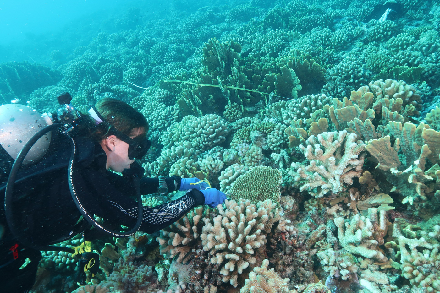
Lauren Howe-Kerr collects a tissue biopsy of the branching coral, Acropora hyacinthus, off the coast of Mo’orea, French Polynesia for genomic preservation and microscopy imaging.
In 2021, a team led by researchers from the National Wildlife Health Center Field Station in Honolulu published a series of images captured using a transmission electron microscope that showed filamentous virus-like particles (VLPs) in dinoflagellates sampled from corals afflicted by SCTLD. The finding led many to ask whether filamentous viruses caused the disease. “People really began to look for different lines of evidence to see whether or not this was true,” Correa said.
Howe-Kerr, who also conducted her PhD research in Correa’s lab formerly at Rice, had previously worked to compile a separate database of more than 700 dinoflagellate cell samples from healthy and bleached coral colonies collected between March 2018 and August 2019. Those samples were taken from reefs off the coast of Mo’orea, French Polynesia, then imaged using a transmission electron microscope. According to Howe-Kerr, the 2021 publication, which included images from corals in Florida, helped members of Correa’s lab correctly identify filamentous VLPs in their images.
“It was wild to identify the VLPs in our images,” she said. “At the time, we thought these structures weren’t part of the normal cell machinery, but they were so common and abundant that we weren’t quite sure they were viruses.”
Navigating uncharted waters
Researchers in Correa’s lab identified filamentous VLPs in images of healthy and bleached coral samples collected in the South Pacific island of Mo’orea even though SCTLD has not been reported in the Pacific Ocean basin. The VLPs were most abundant in dinoflagellates of bleached corals sampled during a marine heatwave, suggesting that high temperatures may worsen these viral infections. They also documented potential filamentous viruses in dinoflagellate cells expelled from coral colonies. The movement of these expelled cells in the water on the reef may be one way these microorganisms are transmitted from one coral colony to another.
“All this suggests that these viruses might actually be widespread in coral dinoflagellates around the world,” explained Howe-Kerr. “This tells us that they are not solely associated with SCTLD and that we need to take a much closer look at what these viruses are doing.”
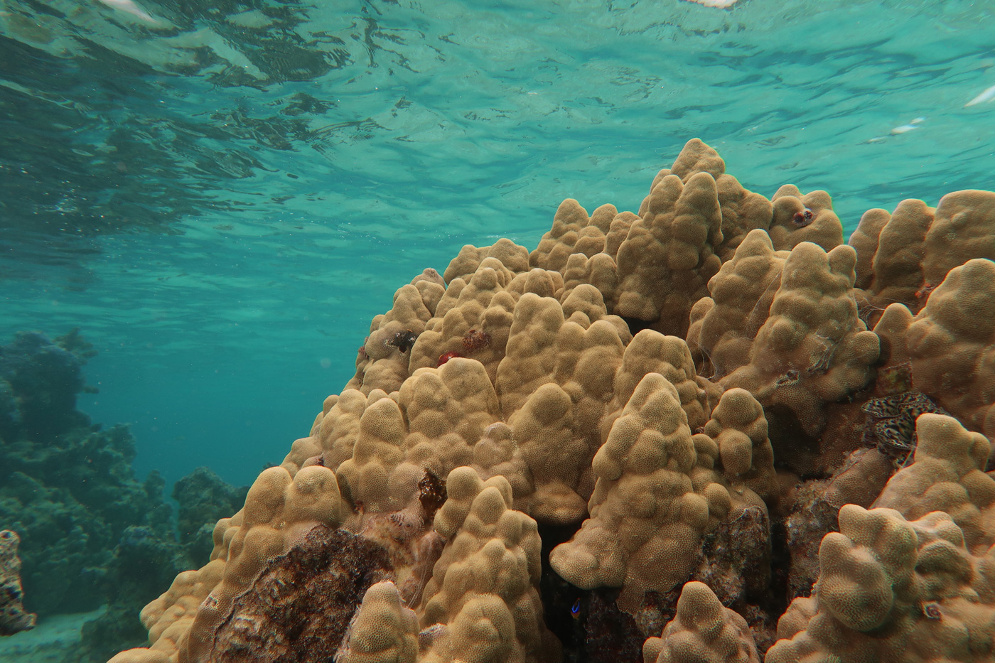
A large mounding coral, Porites cf. lobata, in the back reef waters of Mo’orea, French Polynesia.
Correa, who has studied coral reef virology for the past 14 years, said more research is needed to understand better how viruses and microorganisms affect coral colonies' overall health and function. To advance that goal, her lab is now collaborating with scientists at Oregon State University and the Smithsonian at the University of California’s Gump Research Station to deep sequence corals and other reef organisms—including their resident viruses—on Mo’orea.
“This work will create an unprecedented dataset with a lot of information,” Correa said. “We will likely identify additional viruses that we currently don’t know are found on reefs, and doing that will inherently generate more questions about their function.”
Additional co-authors include Anna Knochel, Matthew Meyer, Carly Karrick, and Alex Veglia of Rice University; Oregon State University professors Andrew Thurber and Rebecca Vega Thurber; George Mason University PhD student Jordan Sims; and Carsten Grupstra, a postdoctoral researcher at Boston University. Funding was provided by the National Science Foundation and the Gordon and Betty Moore Foundation.